Revolutionary Spectral CT Technology Redefines Cardiovascular Diagnostics and Early Cancer Detection at Thang Long Lac Viet International Hospital

Modern medicine has long grappled with the physical boundaries of conventional computed tomography (CT) imaging. Despite decades of technological refinement, traditional CT scanners remain constrained by persistent artifacts: bright streaks generated by high-density metallic components such as surgical stents and artificial joints, the optical obscuration of delicate soft tissues, and the cumulative risk associated with ionizing radiation exposure. These technical constraints have historically acted as invisible barriers, shielding subtle yet lethal pathologies from early detection and precise clinical evaluation.
However, a paradigm shift is underway in diagnostic radiology. At the forefront of this transformation is the deployment of advanced dual-layer spectral CT technology—specifically systems equipped with 256 physical detector rows capable of simultaneous 512-slice reconstruction—which transcends traditional morphological imaging. By moving beyond simple structural visualization, these next-generation scanners can accurately analyze material composition and tissue perfusion at the sub-millimeter level. This breakthrough capability has recently been validated in clinical settings through complex cardiovascular and oncological cases at Thang Long Lac Viet International Hospital, setting a new benchmark for diagnostic precision in modern healthcare.
Overcoming Metallic Artifacts in Complex Cardiac Stenting
The diagnostic challenges of evaluating patients with pre-existing metallic implants have vexed radiologists and cardiologists for decades. When X-ray photons strike the high-density metal framework of a coronary stent, beam hardening and photon starvation occur, scattering the radiation and generating severe streak artifacts. These visual distortions often completely obscure the vascular lumen inside the stent, rendering follow-up assessments unreliable and complicating the detection of in-stent restenosis.
This clinical hurdle is starkly illustrated by the case of Dr. N.V.X., a 48-year-old physician from Hanoi with a complex cardiovascular history. Having previously undergone percutaneous coronary intervention (PCI) to place dual stents in high-risk anatomical locations—specifically the left main coronary artery and the left anterior descending branch—Dr. X. remained completely asymptomatic, reporting no episodes of chest pain. Fully aware of the silent risk of restenosis, he rigorously adhered to routine computed tomography follow-up schedules. Yet, due to photon scatter from the metallic stent struts, all previous conventional CT scans yielded false-positive or inconclusive results, masked by artifacts that veiled the vessel lumen.
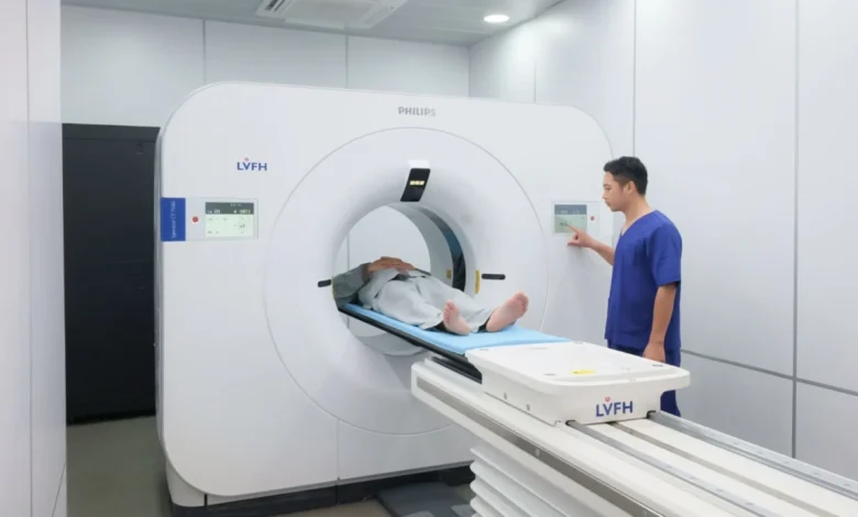
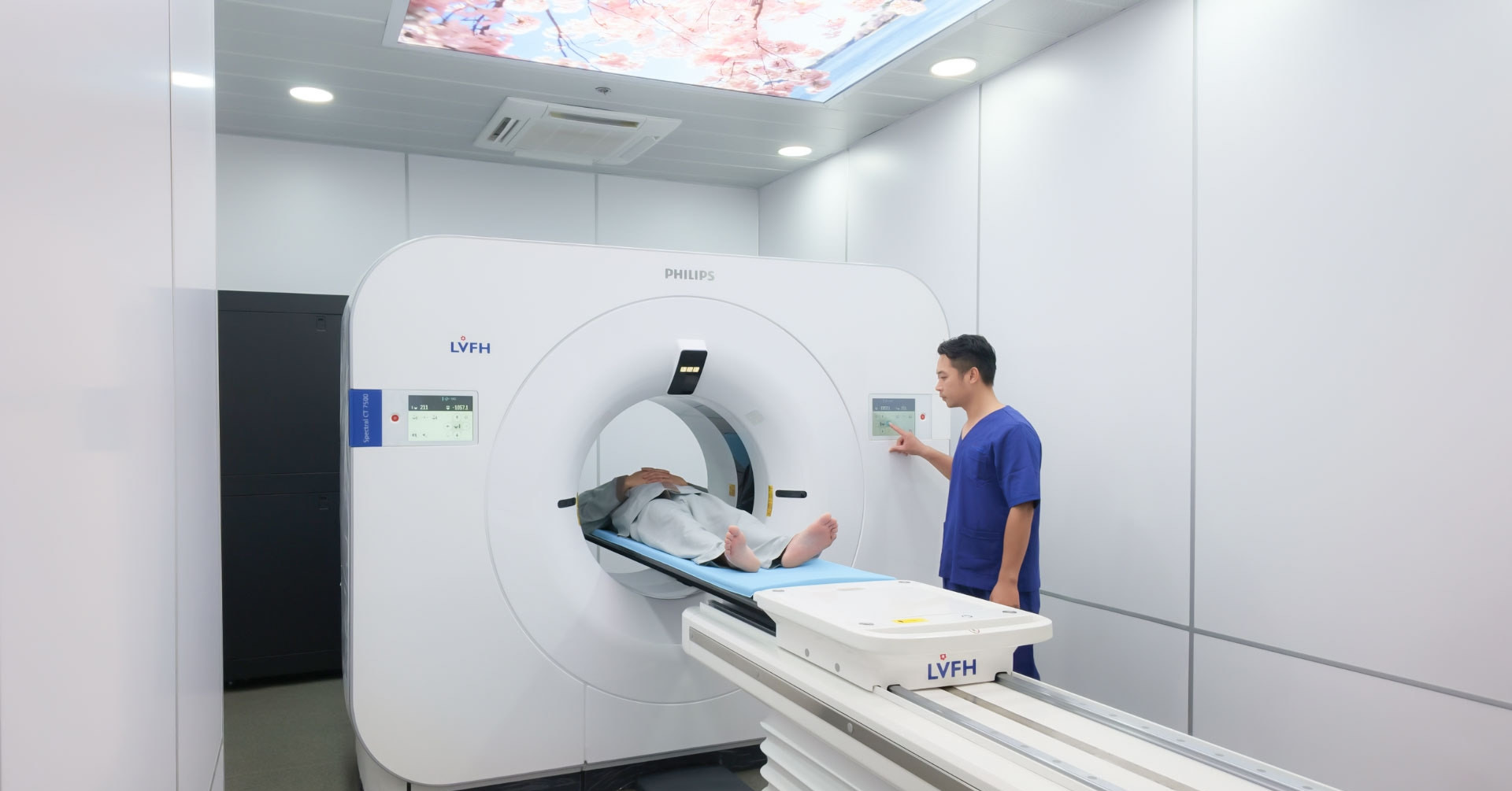
The diagnostic impasse was definitively broken when Dr. X. underwent a comprehensive evaluation utilizing the Philips Spectral CT 7500 scanner at Thang Long Lac Viet International Hospital. The system’s spectral imaging capabilities instantly neutralized the metal artifacts, laying bare the high-definition architecture of the stent lumen. The scan revealed significant luminal narrowing immediately distal to the stent in the left main coronary artery, driven by vulnerable plaque reformation.
Assisted during the multidisciplinary case review by Associate Professor, People’s Doctor, Meritorious Doctor Thai Khac Chau, specialists evaluated the findings. "Metallic stent frameworks have long posed a formidable challenge to diagnostic imaging because scatter artifacts obscure nearly 100% of the vascular lumen," noted Assoc. Prof. Dr. Chau. "Advanced 512-slice spectral CT systems penetrate through metal to accurately assess luminal patency while simultaneously generating myocardial perfusion maps to detect subclinical micro-infarctions that conventional imaging fails to observe."
Promptly managed following these findings, Dr. X. received precise percutaneous intervention, successfully mitigating an impending cardiac event before symptoms could manifest.

Incidental Oncology: Uncovering Malignancy During Cardiac Scans
Beyond cardiovascular applications, the broad screening capabilities and high-resolution material decomposition of spectral CT technology have transformed diagnostic protocols for multi-system evaluations. This dual-utility approach was further demonstrated in the case of P.S.H., a 45-year-old patient from Son La province.
Patient H. presented for medical evaluation following the incidental discovery of a small pulmonary nodule, measuring approximately 7.5 millimeters, located in the middle lobe of the right lung. To comprehensively survey the entire thorax and coronary tree, attending physicians ordered a specialized scan using the Spectral CT 7500 platform, applying advanced respiratory-gated protocols to expand the scan field across the entire chest using a single, optimized dose of iodinated contrast media.
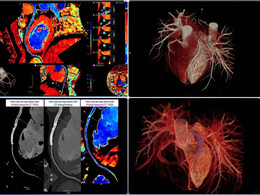
Utilizing iodine quantification maps—an exclusive capability of spectral CT—the perfusion-weighted imaging data immediately highlighted a hyper-enhancing pulmonary nodule within the right lung, showing abnormally high iodine uptake. This pronounced focal hyper-enhancement served as a critical red flag indicating abnormal neo-angiogenesis, a hallmark of malignant tumors that is frequently obscured or overlooked entirely during conventional multi-slice CT examinations.
Without requiring a secondary contrast injection or subjecting the patient to additional ionizing radiation exposure, patient H. received a comprehensive assessment of both her cardiovascular health and an early-stage malignancy. She was immediately referred for targeted 3D-assisted ultrasound and tissue biopsy, opening a clear pathway toward curative intervention and optimal long-term prognosis.
Technological Pillars of Next-Generation Spectral Imaging
The clinical efficacy of advanced dual-layer spectral CT stems from fundamentally redesigned hardware and software architecture. Operating at rapid gantry rotation speeds of one second or less for combined neuro-vascular and cardiac surveys, and under two seconds for whole-body imaging, these systems achieve exceptional temporal resolution. This speed prevents motion artifacts while capturing micro-structures approaching sub-millimeter scales.
Concurrently, integrated advanced dose-reduction algorithms ensure patient safety by minimizing both radiation exposure and contrast agent volume. By capturing high-energy and low-energy photon data simultaneously through dual-layer detector technology, the scanner acquires spectral information on every patient, every scan, without requiring protocol pre-selection. This eliminates the need for repeat scans when unexpected findings emerge, streamlining clinical workflows and reducing patient burden.
The integration of these cutting-edge platforms by pioneering medical centers marks a significant evolution in preventive medicine. Patients are no longer evaluated through isolated diagnostic windows; instead, they benefit from comprehensive, multi-system analyses driven by top-tier clinical expertise and advanced imaging technology.
Implications for the Future of Diagnostic Medicine
The successful clinical deployment of high-slice spectral CT systems establishes a new paradigm for early disease detection, particularly in complex populations with prior interventions or multi-morbidities. By overcoming historical physical limitations such as metallic scatter and low soft-tissue contrast, advanced imaging empowers clinicians to intervene proactively rather than reactively.
As healthcare institutions continue to invest in transformative technologies, the standard for patient care rises correspondingly. The integration of advanced diagnostic hardware with multidisciplinary clinical expertise safeguards public health by identifying life-threatening conditions at their earliest, most treatable stages. This technological evolution underscores a broader commitment within modern medicine: delivering precise, safe, and comprehensive care that redefines what is possible in clinical diagnostics.