
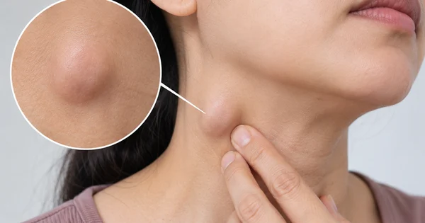
The appearance of a lump in the neck is a clinical phenomenon that frequently causes significant anxiety for patients, often triggering an immediate fear of malignancy. However, from a medical standpoint, the vast majority of neck lumps are not indicators of cancer. Instead, they are typically the body’s localized immune response to an underlying infection or inflammation. Understanding the anatomy, physiology, and pathology of the lymphatic system is essential for patients to distinguish between benign reactions and conditions that require urgent medical intervention.
The Physiology of the Lymphatic System and Neck Lumps
The lymphatic system is a critical component of the human immune response, acting as a drainage and filtration network that transports lymph fluid—a clear fluid containing white blood cells—throughout the body. Lymph nodes, which are small, bean-shaped structures, act as regional outposts where the immune system filters out pathogens such as bacteria, viruses, and cellular debris.
The head and neck region contains a dense concentration of these nodes, making it the most common site for noticeable swelling. According to medical experts at the Mayo Clinic, when a person suffers from a common cold, a sore throat, tonsillitis, an ear infection, or dental abscesses, the neighboring lymph nodes often enlarge as they work to neutralize the invading microbes. This process is a sign of a functioning immune system rather than a disease process itself.
Clinical Chronology: From Infection to Resolution
The typical timeline of a reactive lymph node follows the progression of the underlying infection. In most cases, a patient will notice one or more small, firm lumps shortly after the onset of symptoms like a sore throat or fever.
- Initial Phase (Days 1–7): As the primary infection peaks, the lymph nodes begin to swell. During this phase, the nodes may feel tender or painful when touched. This discomfort is often a result of rapid cell proliferation within the node as it fights the infection.
- Resolution Phase (Weeks 1–3): As the immune system successfully manages the primary infection—either naturally or with the aid of antibiotics—the inflammatory stimulus is removed. The lymph nodes should gradually decrease in size and return to their baseline state.
- Observation Phase (Weeks 4+): If the lump persists beyond four weeks, or if it continues to enlarge despite the resolution of the initial illness, the clinical picture shifts. This is the stage where medical professionals advise a transition from observation to diagnostic investigation.
Distinguishing Benign Reactions from Malignancy
While it is crucial to remain calm, it is equally important not to ignore persistent symptoms. The fear of lymphoma or metastatic cancer is common, yet these represent a minority of cases. Malignancy-related lymphadenopathy generally exhibits different clinical characteristics than reactive nodes.
According to data from the Medical News Today health portal, physicians evaluate neck masses based on several key diagnostic markers:
- Consistency: Benign, reactive nodes are often soft or rubbery and movable beneath the skin. Malignant nodes, conversely, may feel hard, fixed in place, or painless.
- Growth Pattern: Nodes that continue to grow in size over weeks or months are statistically more concerning than those that fluctuate in size or remain stable.
- Systemic Symptoms: The presence of "B symptoms"—which include unexplained weight loss, drenching night sweats, and persistent fevers—are significant clinical red flags that necessitate immediate imaging, such as an ultrasound or CT scan, and potentially a biopsy.
The Role of Diagnostic Imaging and Biopsy
In a clinical setting, if a patient presents with a neck lump that does not resolve within the expected timeframe, the diagnostic protocol is systematic. Initially, a physician will perform a physical examination to determine the location, mobility, and tenderness of the node.
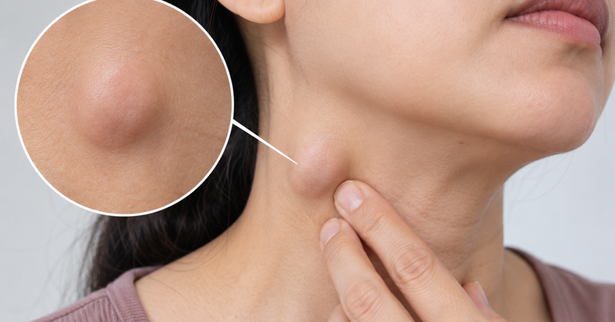
If suspicion of a more serious condition remains, imaging is the next logical step. Ultrasound is often the preferred first-line diagnostic tool because it is non-invasive and can provide high-resolution images of the node’s internal architecture, blood flow, and margins. If the ultrasound reveals suspicious features, a fine-needle aspiration (FNA) biopsy may be performed to extract cells for laboratory analysis. This provides a definitive diagnosis, allowing clinicians to distinguish between reactive hyperplasia, granulomatous infections like tuberculosis, or various forms of lymphoma.
Broad Implications: When to Seek Medical Attention
The psychological impact of discovering a neck lump cannot be overstated. Health anxiety often leads patients to perform frequent self-examinations, which can ironically exacerbate the condition. Medical experts explicitly advise against the habit of repeatedly pressing, squeezing, or manipulating the lump. Such tactile stimulation can cause further irritation to the lymphatic tissue, leading to localized inflammation and potentially masking the true clinical progression of the node.
It is important for the public to recognize that the absence of pain does not necessarily mean a lump is benign. Conversely, painful lumps are more frequently associated with active infections. The most reliable indicator for when to seek professional help is the persistence of the mass. If a lump remains present for more than two to four weeks, regardless of whether it is painful or not, it warrants a clinical evaluation.
Public Health Perspective
From a broader public health perspective, the rise in awareness regarding lymphatic health has led to earlier detection of various conditions. While the internet has empowered patients with information, it has also increased the prevalence of "cyberchondria," where patients self-diagnose based on incomplete data.
Professional medical organizations emphasize that there is no substitute for a physical examination. Factors such as the patient’s age, medical history, smoking status, and exposure to environmental toxins are critical variables that a doctor considers when assessing a neck mass. For instance, in older patients, a persistent neck mass is treated with a higher index of suspicion for squamous cell carcinoma of the head and neck, especially if there is a history of tobacco or alcohol use.
Conclusion and Recommendations
The appearance of a lump in the neck is a common, often benign, and temporary reaction to the body’s ongoing immune activities. It is a sign that the body is working to protect itself. However, the threshold for seeking medical advice should be clearly defined:
- Monitor: Observe the lump for two weeks after the initial infection has cleared.
- Avoid Manipulation: Do not touch or squeeze the area, as this can irritate the tissue.
- Consult: If the lump persists beyond four weeks, grows in size, becomes fixed to surrounding tissues, or is accompanied by unexplained weight loss or night sweats, schedule an appointment with a primary care physician or an otolaryngologist (ENT specialist).
By maintaining a balanced perspective and adhering to these professional guidelines, patients can effectively manage their health, reduce unnecessary anxiety, and ensure that any potentially serious condition is addressed with the appropriate medical urgency. In the modern era of medicine, early detection remains the most effective tool in managing lymphatic health, turning what could be a source of fear into a manageable aspect of routine healthcare.
Related posts: