
Understanding Thymic Carcinoma: Clinical Insights, Diagnostic Challenges, and Current Treatment Paradigms

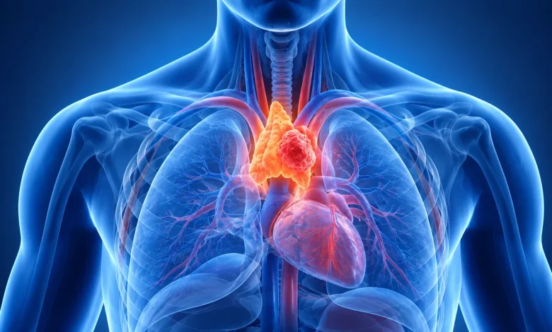
Thymic carcinoma is a rare and aggressive malignancy originating from the epithelial cells of the thymus, a specialized organ located in the upper anterior mediastinum, playing a critical role in the development of the immune system’s T-lymphocytes. While the thymus is essential during childhood for "training" immune cells to distinguish between self and non-self, its physiological significance wanes in adulthood, leaving the organ susceptible to rare oncological transformations. Unlike thymomas, which are typically indolent and encapsulated, thymic carcinomas are characterized by rapid cellular proliferation, a high propensity for local invasion into surrounding structures like the pericardium and great vessels, and a significant risk of distant metastasis.
Defining the Pathology and Classification
The clinical classification of tumors arising from the thymus is generally grouped under Thymic Epithelial Tumors (TETs). Within this spectrum, thymic carcinoma represents a distinct and more lethal subtype compared to thymomas. Pathologically, these tumors are classified based on the World Health Organization (WHO) criteria, which categorize them by the morphological features of the epithelial cells.
Thymic carcinomas, specifically categorized as Type C thymic epithelial tumors, account for approximately 20% of all thymic malignancies. Unlike the more common thymoma, which often presents with an intact capsule, thymic carcinoma exhibits clear cytological atypia and lacks the characteristic immature T-cell infiltrate found in lower-grade thymic tumors. The aggressive nature of these cells means they often outgrow their local environment, necessitating a highly specialized approach to diagnosis and intervention.
The Diagnostic Challenge: Symptoms and Silent Progression
One of the most profound challenges in the clinical management of thymic carcinoma is its tendency to remain asymptomatic in its early stages. Because the tumor resides in the mediastinum—a space occupied by the heart, the trachea, the esophagus, and major blood vessels—symptoms often only manifest when the mass has reached a size sufficient to exert mechanical pressure on these vital structures.
Common clinical presentations include persistent cough, chest pain, dyspnea (shortness of breath), and dysphagia (difficulty swallowing). In some instances, patients may present with symptoms related to superior vena cava syndrome, where the tumor compresses the primary vein returning blood from the upper body, leading to facial swelling and venous distension.
Furthermore, thymic carcinoma is occasionally associated with paraneoplastic syndromes, most notably myasthenia gravis. This autoimmune condition occurs when the body’s immune system mistakenly attacks the communication junction between nerves and muscles. Symptoms include ptosis (drooping eyelids), diplopia (double vision), limb weakness, and, in severe cases, respiratory crisis. While myasthenia gravis is more classically associated with thymomas, its presence in a patient with a mediastinal mass should immediately raise suspicion of a thymic epithelial pathology. Because these symptoms are non-specific and mimic common respiratory ailments, many cases are discovered incidentally during routine X-rays or CT scans performed for unrelated conditions.
Epidemiological Context and Etiological Ambiguity
The medical community continues to grapple with the underlying causes of thymic carcinoma. Unlike lung cancer, where smoking is a well-established driver, or liver cancer, where viral hepatitis plays a central role, the etiology of thymic carcinoma remains largely elusive. Researchers have not yet identified a specific environmental exposure or lifestyle factor that reliably predicts the development of this malignancy.
Current scientific consensus points toward a complex interplay of genetic mutations occurring within the thymic epithelial cells. These somatic mutations cause the cells to bypass standard apoptotic pathways—the programmed cell death that prevents uncontrolled growth—leading to the formation of a persistent, malignant mass. While some research has investigated familial genetic predispositions, the majority of thymic carcinoma cases appear to be sporadic. This lack of a clear "risk profile" complicates early screening efforts, as there is no specific high-risk population to monitor.
Chronology of Clinical Management
The diagnostic journey for a patient suspected of having thymic carcinoma typically follows a structured protocol:
- Initial Detection: Often through incidental radiological imaging (X-ray or CT) showing a widening of the mediastinum.
- Advanced Imaging: Enhanced CT or MRI scans are utilized to map the tumor’s precise dimensions and its proximity to the heart and major blood vessels.
- Tissue Assessment: In cases where the tumor is clearly resectable, surgeons may proceed directly to surgery. In more complex or advanced cases, a CT-guided biopsy may be required to confirm the histological subtype before finalizing a treatment plan.
- Staging: Utilizing the TNM system—which measures Tumor size (T), Lymph Node involvement (N), and Metastasis (M)—clinicians determine the severity of the disease. This stage directly dictates whether the patient is a candidate for curative surgery or requires systemic therapy.
The Multimodal Treatment Paradigm
Because of the rarity of thymic carcinoma, treatment decisions are rarely standardized and are best managed by multidisciplinary tumor boards consisting of thoracic surgeons, medical oncologists, and radiation oncologists.
Surgery: The Gold Standard
For patients with localized disease, surgical resection is the primary and most effective treatment. The objective is to achieve an "R0 resection," meaning the tumor is removed in its entirety with negative margins. If the tumor has invaded adjacent structures, surgeons may perform an "extended" resection, which involves removing portions of the pericardium, pleura, or even major vessels to ensure the cancer is completely cleared.
Radiation Therapy
Radiation is frequently employed as an adjuvant (post-surgical) treatment to eradicate any microscopic residual disease, thereby reducing the risk of local recurrence. In cases where the tumor is deemed unresectable due to its involvement with critical structures, high-energy radiation may be used as the primary modality to control growth and alleviate obstructive symptoms.
Chemotherapy
Systemic chemotherapy is a cornerstone for patients with advanced-stage thymic carcinoma or those whose tumors have metastasized. Chemotherapeutic agents are delivered intravenously to circulate throughout the body, targeting rapidly dividing cancer cells. In some clinical scenarios, "neoadjuvant" chemotherapy is administered before surgery to shrink the tumor, potentially making a previously inoperable case amenable to surgical intervention.
Implications and Future Directions
The prognosis for thymic carcinoma is historically guarded due to the late stage at which many patients are diagnosed. However, advancements in oncology are shifting the landscape. The move toward personalized medicine, specifically the use of genomic profiling, is helping researchers understand the molecular drivers of thymic carcinoma. By identifying specific genetic signatures, clinicians are beginning to explore targeted therapies and immunotherapies that may offer more efficacy than traditional cytotoxic chemotherapy.
From a public health perspective, the implications are clear: increased awareness of persistent respiratory symptoms—even in the absence of a smoking history—is essential. While rare, thymic carcinoma highlights the necessity of investigating unexplained chest pain or difficulty breathing with high-resolution imaging.
The integration of centralized registries and international clinical trials is also critical. Because individual hospitals may see only a handful of these cases per year, global collaboration is the only way to generate the data necessary to refine treatment protocols. As we look toward the future, the goal is to shift from reactive, symptomatic management to early, precise detection, ultimately improving the survival rates and quality of life for patients diagnosed with this complex malignancy. For patients and families navigating this diagnosis, the most critical step remains seeking care at high-volume specialized centers where the nuances of this rare tumor are understood and managed through a unified, multidisciplinary effort.